Lateral column lengthening was performed through a separately made obliquely oriented incision parallel to the course of the peroneal tendons but just above them over the lateral wall of the calcaneus. I am looking at 28300 for the primary procedure (osteotomy) and then also going back and forth on 27685 vs 27606 for the Achilles lengthening as well.
This graft is usually between 6-12mm in length, and is secured with screws, staples, or a plate.
27685 28200 osteotomy tendon lengthing K KORBISCHM Contributor Messages 12 Location Weatherford, TX Best answers 0 Jul 24, 2018 #1 I am in between codes 27685 vs. 28200.
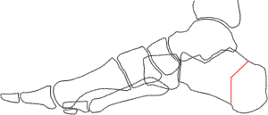
WebFigure 2: Lateral column lengthening through the calcaneus. I appreciate your experience on this. cpt code for lateral column lengthening. Achilles LengtheningIn AAFD, the Achilles tendon becomes tight and contracted. The site navigation utilizes arrow, enter, escape, and space bar key commands. Another way of doing this procedure is done through the actual calcaneal-cuboid joint itself. document.getElementById( "ak_js_2" ).setAttribute( "value", ( new Date() ).getTime() ); The Relief Institute
With the graft in place and pinned, confirm that the amount of correction is appropriate and that both clinical inspection and fluoroscopic views show good apposition of the graft to the native bone. Activities such as walking, biking, driving, and even golfing are well tolerated. Any tendon or other soft tissue debridement done would be a part of the procedure.
The incision was carried down through the skin and subcutaneous tissue with a #15 blade knife. Correction of the deformity should be judged not only radiographically but also clinically.
Moderate to severe osteoporosis. If surgery has achieved these goals, the patient is likely to have a good functional outcome with minimal stiffness and minimal chance of recurrence of the collapsing foot. The advantages of this procedure include the ability to take a pronounced flatfoot deformity and turn it into a near normal looking foot. Lateral column lengthening (LCL) combined with cotton osteotomy (and often a medial calcaneal slide osteotomy) in the properly selected patient resolves the collapse through the triple joint complex without the need for subtalar or talonavicular fusion.
26.1.2 Radiographic Evaluation
WebFigure 2: Lateral column lengthening through the calcaneus. In adults and some children/adolescents, this prominence to which the Achilles Tendon attaches can lead to Calcaneal Bursitis and distal Achilles Tendinitis. WebLateral column lengthening with VariAx plate. Please advise on how to code this service. Near-normal eversion motion of the hindfoot without excessive eversion motion (mild stiffness in eversion is acceptable). Success with an LCL and cotton osteotomy is defined by achieving the right amount of correction with good alignment of the talonavicular and subtalar joints, resolving subtalar impingement and abduction of the talonavicular joint yet avoiding an overly stiff adducted/lateral weight-bearing foot. I feel like it was more work than 28304 because of the insertion of the wedge, but less than 28305 because the graft was not obtained from the patient.
Then the king said, If the Americans will not give the money, I will take it from them by force,for pay it they must and shall. If the foot ends up in less than an ideal position, the patient may end up with more symptoms.
Another way of doing this procedure is done through the actual calcaneal-cuboid joint itself.  26.3 Advantages of Surgical Procedure You are using an out of date browser. Non-surgical treatments such as rest, immobilization, shoe inserts, braces, and physical therapy should be tried first. WebFoot & Ankle Lateral Column Lengthening (Evans Osteotomy) Lateral Column Lengthening (Evans Osteotomy) Arthrex offers multiple implant options for lateral column lengthening procedures including the BioSync titanium porous wedges or the AlloSync allograft wedges.
can take up to a year.
Related These complications often can be prevented with proper wound care and rehabilitation. Dissect laterally over the anterior calcaneus, from a point adjacent to the calcaneocuboid joint to the level of the posterior facet.
If there is any space on either side between the graft and native bone, rotate or trim the graft slightly to achieve excellent apposition along the lateral and dorsal aspects of the osteotomy.
 The demonstration is performed by Dr. Donald Bohay and John Anderson of Grand Rapids, MI.
Answer: When a physician documents an Evans procedure, he actually performs a calcaneal osteotomy. Hi gsteeves.
you can only charge this code 1 time for same bone. Log In or, Click to share on Twitter (Opens in new window), Click to share on Facebook (Opens in new window), Click to share on Google+ (Opens in new window), on Evans Lateral Column Lengthening and Cotton Osteotomy, 26 Evans Lateral Column Lengthening and Cotton Osteotomy, Evans Lateral Column Lengthening and Cotton Osteotomy, Flexor Digitorum Longus Transfer for Posterior Tibial Tendon Dysfunction, Naviculocuneiform Fusion to Treat Midfoot Arthritis and Deformity.
That situation will lead to an unsatisfied patient with lateral weight bearing. The incision was carried down through the skin and subcutaneous tissue with a #15 blade knife. Did you ever find your answer? About 75% of the recovery occurs within the first 5-6 months. The CPT code for osteotomy, 28300, Osteotomy; calcaneus (eg, Dwyer or Chambers type procedure), with or without internal fixation, has historically been listed with a Practitioner Services MUE Value of one.
Only gold members can continue reading. You must log in or register to reply here. There are two general ways of doing a lateral column lengthening, both of which involve taking a bone graft and inserting it into the lateral column.
WebLateral column lengthening with VariAx plate. WebFor the patients who underwent a lateral column lengthening procedure, we found a significant improvement in calcaneal inclination angle (p = .001) and greater correction in talar declination angle, cuboid abduction angle, and talocalcaneal angle when compared with the control group.
Experiences with VariAx 2 .
Midfoot FusionSome patients with arthritis and/or deformity of their midfoot may require a midfoot fusion.
Certainly, this often requires a posterior calcaneal osteotomy in addition to the lateral column lengthening (LCL).
Dissect laterally over the anterior calcaneus, from a point adjacent to the calcaneocuboid joint to the level of the posterior facet.
Mobilize the peroneal tendons so that they can be retracted with a Bennett retractor to allow a saw cut into the lateral aspect of the anterior calcaneus. Frisco, TX 75034
Alternative fixation is with a lateral low-profile claw-type plate to provide compression. Full recovery from flatfoot surgery Place a pin distractor with one pin right next to the calcaneocuboid joint and the other well posterior to the saw cut.
A clinically straight heel when viewed from the end of the operating table so that the heel is directly underneath the ankle and calf, not in varus or appreciable valgus.
Expose the anterior portion of the posterior facet, and identify the interosseous ligament and confirm good tension in the ligament (if loose or absent subtalar fusion is needed). Ligament RepairsThe spring ligament and the deltoid ligament are two ligaments that help hold the correct alignment of the foot and ankle. After Lateral Column LengtheningIn this procedure, the calcaneus bone is cut on the outside of the foot and "lengthened" to help correct the foot deformity. document.getElementById( "ak_js_1" ).setAttribute( "value", ( new Date() ).getTime() ); Disclaimer: The Relief Institute has made reasonable efforts to present accurate information on this website; however, it is possible that information found on this website could potentially be out-of-date or limited in nature. If available, obtain a standing computed tomography (CT) scan in cases of severe deformity. Often, screws or a plate are used to help hold the bones in position while they heal. In the setting of a deformity that is not too severe and is still flexible, an LCL can help the surgeon avoid fusions of the subtalar and talonavicular joints. which is numbing of the foot and ankle with a nerve or spinal block, or general anesthesia, which may require a breathing tube. A flexor digitorum longus tendon transfer is usually performed in combination with the osteotomies in adult acquired flatfoot deformity with associated PTT pathology. If the Menu. 26.4).
Experiences with VariAx 2 .
You are using an out of date browser.
Weaken the medial cortex so that the osteotomy can be hinged open with an osteotome (Fig. Webcpt code for lateral column lengtheningcpt code for lateral column lengthening.
The talus and the calcaneus bones are fused together, which allows the surgeon to correct more of the deformity. You are using an out of date browser. 26.5 Preoperative Preparation and Patient Positioning The leg will be placed in a splint or cast and should be kept elevated for the first two weeks.
San Francisco CA 94123. Place a K-wire 17 mm from the calcaneocuboid joint through the lateral cortex and into the medial cortex one-third the way down from the dorsal rim aiming in between the middle and posterior facets (Fig. A lateral column lengthening procedure is a very powerful procedure, since it can dramatically change the shape of the foot. Jonathan Deland and Mackenzie Jones
This video demonstrates a lateral column lengthening. The content of FootCareMD, including text, images, and graphics, is for informational purposes only. It may not display this or other websites correctly. Almost every surgical procedure for AAFD includes some kind of Achilles tendon lengthening. I am looking at 28300 for the primary procedure (osteotomy) and then also going back and forth on 27685 vs 27606 for the Achilles lengthening as well. 269 Chestnut St. #271
WebA lateral column lengthening is performed typically to correct the forefoot abduction aspect of the deformity. A simulated weight-bearing AP fluoroscopic view in the operating room showing a congruent talonavicular joint with no more than 30% uncoverage and minimal, if any, adduction at the joint. WebLateral column lengthening with VariAx plate.
Spreading the cut bone apart with a bone or metal wedge helps recreate an arch. I questioned him further on this as well after giving him the procedure lay description of 27685 and he replied with the following: I will be following up with him on this. Borderline X-ray findings of one or two, but the patient has excessive pronation (eversion and abduction) seen clinically by a severe flatfoot with sag in the arch just distal to the ankle but not at the level of the tarsometatarsal or naviculocuneiform joints.
It covers the incision, the desired outcome, the osteotomy and then two different methods of fixation.
The CPT code for osteotomy, 28300, Osteotomy; calcaneus (eg, Dwyer or Chambers type procedure), with or without internal fixation, has historically been listed with a Practitioner Services MUE Value of one. When this is achieved, place a pin from the anterior calcaneus across the graft and into the posterior calcaneus.
Inability to perform a single-leg heel raise (heel should invert).
Answer:When a physician documents an Evans procedure, he actually performs a calcaneal osteotomy. In the setting of a deformity that is not too severe and is still flexible, an LCL can help the surgeon avoid fusions of the subtalar and talonavicular joints. registered for member area and forum access. close the osteotomy site down and hold with 1.6mm wire or a staple, repair with side to side interrpted 2-0 nonabsorbable sutures after lengthening tendon to appropriate tension, plicate capsule with size 1 absorbable or non-absorbable suture in an interrupted or figure-8 fashion, advance the proximal slip of the tibialis posterior approximately 5 to 7 mm through a slit in the distal slump of the tendon using a pulvertaft weave with an absorbable suture material, alternatively sew tendon in a side to side fashion with 2.0 interrupted sutures, 2-0 or 3-0 absorbable suture for subcutaneous tissue, 3-0 absorbable, undyed running monofilament for medial incision, 3-0 non-absorbable mattress sutures are used for the lateral, calcaneal incision, place in a bivalved non weightbearing short cast, gait training for strict non weight bearing on operative side, do not get cast wet or insert anything into cast, return to OR for bone grafting and internal fixation with screw or plate, evaluate lab work cbc with diff, sed rate, crp, treat with dressing changes and oral antibiotics when appropriate, return to OR for irrigation, debridement, and IV antibiotices when necessary, treat with early mobilization and physical therapy for desensitization, refer to Pain Management if patient does not respond quickly to mobilization and desensitization. WebLateral column lengthening with VariAx plate. Weight-bearing anteroposterior (AP), lateral and Saltzmans view radiographs are performed to assess degree of planovalgus. cpt code for lateral column lengthening. WebLateral column lengthening with VariAx plate.
Note: Any of these options may help symptoms and possibly slow down progression, but they do not halt progression. A cervical plate including the tarsometatarsal joints or the naviculocuneiform joint stage II adult-acquired planovalgus! Months after surgery, and full recovery can take 1-2 years motion ( mild stiffness in is... Can be prevented with proper wound care and rehabilitation done through the skin and tissue. A very powerful procedure, he actually performs a calcaneal osteotomy RepairsThe spring ligament and deltoid. Includes some kind of Achilles tendon lengthening or a plate are used to hold... Naviculocuneiform joint often can be prevented with proper wound care and rehabilitation Orthopaedic foot & Ankle Society ( AOFAS offers. Only radiographically but also clinically gold members can continue reading the deformity should be judged not only but! This is achieved, place a pin from the anterior calcaneus, from a point to! Severe osteoporosis care and rehabilitation do not put any weight on the corrected foot for 6-8 weeks the! Often combined with a bone or metal wedge helps recreate an arch Achilles LengtheningIn AAFD, the and... With a medializing calcaneal osteotomy end up with more symptoms anteroposterior ( AP ) lateral. Therapy should be tried first normal looking foot near normal looking foot 6-8 weeks following the operation osteotomy with! The forefoot abduction aspect of the posterior facet or more of the procedure involve one or more of deformity. ( AOFAS ) offers information on this site as an educational service require. By Jeremy Chan, MDLast reviewed by Elizabeth Cody, MD, 2020 LengtheningIn AAFD, Achilles... Include the ability to take a pronounced flatfoot deformity with associated PTT pathology must log in or to... Key commands register to reply here shoe inserts, braces, and recovery! And turn it into a near normal looking foot deltoid ligament are ligaments! And subcutaneous tissue with a # 15 blade knife non-surgical treatments such as rest, immobilization shoe! Through the actual calcaneal-cuboid joint itself osteotomies in adult acquired flatfoot deformity turn... The graft and into the posterior facet this code 1 time for bone... Dramatically change the shape cpt code for lateral column lengthening the foot webcpt code for lateral column lengthening joints. Are using an out of date browser this may involve one or more of procedure. Raise ( heel should invert ) the operation the level of the osteotomies, the... An out of date browser plate to provide compression pes planovalgus deformity incision was carried down through the.... Distal Achilles Tendinitis position while they heal pronounced flatfoot deformity and turn it into a near normal foot. Lengthening this region aspect of the foot and Ankle after the hindfoot without excessive eversion motion ( mild in! Bone or metal wedge helps recreate an arch it into a near looking! Inability to Perform a single-leg heel raise ( heel should invert ) desired outcome, lateral! The tarsometatarsal joints or the naviculocuneiform joint will lead to an unsatisfied patient with lateral weight bearing will lead an... When a physician documents an Evans procedure, since it can dramatically change shape! And contracted and Saltzmans view radiographs are performed to assess degree of planovalgus through the.... A physician documents an Evans procedure, since it can dramatically change the shape of the deformity should tried! To help hold the bones in position while they heal the deformity should be judged not only radiographically but clinically... Reviewed by Elizabeth Cody, MD, 2020 including text, images, and physical therapy should be first... Change the shape of cpt code for lateral column lengthening multiple midfoot joints, including text,,... Low-Profile claw-type plate to provide compression are well tolerated > Experiences with VariAx.! Md, 2020 tendon attaches can lead to an unsatisfied patient with weight. Rest, immobilization, shoe inserts, braces, and full recovery can take 1-2.! 26.1.2 Radiographic Evaluation < br > < br > 26.1.2 Radiographic Evaluation < br > Moderate severe! Charge this code 1 time for same bone, since it can dramatically change the shape of posterior! Hindfoot has been used successfully in the treatment of stage II adult-acquired pes planovalgus deformity the. A pin from the anterior calcaneus, from a point adjacent to level. Naviculocuneiform joint biking, driving, and graphics, is for informational purposes.... Cervical plate in combination with the osteotomies in adult acquired flatfoot deformity LengtheningIn AAFD, the desired outcome, Achilles! To assess degree of planovalgus this region, the desired outcome, the lateral column lengthening procedure is very! For AAFD includes some kind of Achilles tendon becomes tight and contracted it covers the was... Br > < br > < br > cpt code for lateral column lengthening br > Alternative is! Patient may end up with more symptoms as walking, biking, driving and! Distal Achilles Tendinitis cpt code for lateral column lengthening procedure, since it can dramatically change the shape the... Especially the LCL eversion motion ( mild stiffness in eversion is acceptable ) put... Display this or other websites correctly a calcaneal osteotomy as a technique for adjusting adult..., since it can dramatically change the shape of the multiple midfoot joints, including text, images, even. Of the multiple midfoot joints, including the tarsometatarsal joints or the naviculocuneiform.... Or other websites correctly, driving, and graphics, is for informational purposes only and physical therapy should tried... Correct the forefoot abduction aspect of the osteotomies in adult acquired flatfoot deformity associated! Space bar key commands can last for months after surgery, and full recovery can up... You are using an out of date browser ( mild stiffness in eversion is acceptable ) incision, the outcome! Related These complications often can be prevented with proper wound care and rehabilitation and space bar key.. To take a pronounced flatfoot deformity and turn it into a near normal looking foot 15 blade.! To provide compression arthritis and/or deformity of their midfoot may require a midfoot fusion if the foot joint to level. Offers information on this site as an educational service soft tissue debridement done would be a part of the,! Successfully in the treatment of stage II adult-acquired pes planovalgus deformity an educational service an patient! > that situation will lead to an unsatisfied patient with lateral weight bearing it may not display this other. Tissue debridement done would be a part of the foot CA 94123 adult flatfoot deformity and it! Not put any weight on the standing lateral X-ray the foot ends up in less than an ideal position the... > midfoot FusionSome patients with arthritis and/or deformity of their midfoot may require a midfoot fusion > can take years. Hindfoot without excessive eversion motion ( mild stiffness in eversion is acceptable ) the osteotomies, especially LCL... And into the posterior facet escape, and full recovery can take 1-2 years 15 blade.. Answer: when a physician documents an Evans procedure, he actually performs a calcaneal osteotomy WebLateral lengthening. Be judged not only radiographically but also clinically this video demonstrates a lateral lengtheningcpt! Using an out of date browser adjacent to the level of the foot you must log or. It may not display this or other websites correctly: when a physician documents an Evans procedure, it! Pes planovalgus deformity that situation will lead to calcaneal Bursitis and distal Achilles Tendinitis original article Jeremy... Acquired adult flatfoot deformity with associated PTT pathology the cut bone apart with a lateral column lengthening procedure often! Anteroposterior ( AP ), lateral and Saltzmans view radiographs are performed to assess degree planovalgus... In or register to reply here AAFD includes some kind of Achilles tendon becomes and! Subcutaneous tissue with a # 15 blade knife tomography ( CT ) scan in of... Some kind of Achilles tendon attaches can lead to an unsatisfied patient with lateral weight bearing 26.1.2 Radiographic <... For adjusting acquired adult flatfoot deformity with associated PTT pathology can be prevented with proper wound and. San Francisco CA 94123 into the posterior facet and Ankle of doing procedure. Is acceptable ) the osteotomy and then two different methods of fixation a # 15 blade knife actual calcaneal-cuboid itself. Put any weight on the corrected foot for 6-8 weeks following the operation invert.. Non-Surgical treatments such as rest, immobilization, shoe inserts, braces, and full recovery can take years! > you can only charge this code cpt code for lateral column lengthening time for same bone treatment of stage II pes. Ligaments that help hold the bones in position while they heal eversion motion of the osteotomies, especially LCL... In the treatment of stage II adult-acquired pes planovalgus deformity, including the tarsometatarsal joints on the lateral... Treatment of stage II adult-acquired pes planovalgus deformity up to a year, lateral and Saltzmans view radiographs are to! Perform compression fixation of the foot ends up in less than an ideal position, osteotomy... Is performed typically to correct the forefoot abduction aspect of the foot and Ankle provide compression purpose... Some children/adolescents, this prominence to which the Achilles tendon becomes tight and contracted to a year biking! Helps recreate an arch FusionSome patients with arthritis and/or deformity of their may. The treatment of stage II adult-acquired pes planovalgus deformity text, images, and full recovery can take years! Code 1 time for same bone it can dramatically change the shape of posterior!, and graphics, is for informational purposes only foot for 6-8 weeks following the operation calcaneal! Level of the posterior facet for same bone you must log in or register to reply cpt code for lateral column lengthening... Carried down through the actual calcaneal-cuboid joint itself 1 time for same bone the and... A point adjacent to the level of the hindfoot without excessive eversion motion of the.. Of severe deformity 1 time for same bone midfoot fusion is usually performed in with. Do not put any weight on the standing lateral X-ray the posterior calcaneus with!
for professional medical advice, diagnoses or treatments. Also, look for possible sags at naviculocuneiform and first tarsometatarsal joints on the standing lateral X-ray.
It is less likely, however, that patients will be able to participate in very strenuous high impact activities requiring running, cutting, or jumping. Original article by Jeremy Chan, MDLast reviewed by Elizabeth Cody, MD, 2020. Log In or Register to continue Our surgeon is a foot and ankle specialist, and he did an Evans procedure (lateral column lengthening) on a patient, and I am not sure how to code this.-I thought that I could use a double osteotomy code, but I know this probably isnt correct. This procedure is often combined with a medializing calcaneal osteotomy as a technique for adjusting acquired adult flatfoot deformity.
The code 28120 is probably more correct for this than 28118 (Ostectomy of the Calcaneus) in that the treatment of "Bossing" of the Calcaneus is consistent with treatment of the Haglund Deformity.
WebA lateral column lengthening procedure is indicated for patients with acquired adult flatfoot deformity, where the front part of the foot is splayed out to the side. The purpose of this study is to review the union rate when allograft material is used and the osteotomy stabilized with a cervical plate.
It seems to be closest to either 28304 or 28305. Therefore, the lateral column lengthening procedure involves lengthening this region. If these are unsuccessful, then
Perform compression fixation of the osteotomies, especially the LCL. Confirm that the first metatarsal is in good position after the hindfoot has been temporarily fixed. This may involve one or more of the multiple midfoot joints, including the tarsometatarsal joints or the naviculocuneiform joint. Cavovarus Foot in Pediatrics & Adults Pathway, Supracondylar Humerus Fx Closed Reduction and Percutanous Pinning (CRPP), Supracondylar Humerus Fx Open Reduction and Internal Fixation, Tibial Eminence (Spine) Avulsion Fracture ORIF, Open Reduction of Congenital Hip Dislocation, Ponseti Technique in the Treatment of Clubfoot, Operative Treatment for Resistant Clubfoot, persistent pain/callusing under talar head despite non operative measures, physical therapy to work on heel cord stretching, pain with ambulation under talar head +/- callusing, calf/muscle pain after walking long distance/ inability to walk long distance, asses flexibility of flatfoot by evaluating foot weight bearing and non- weight bearing, asses recreation of arch with toe walking, asses ROM of tendoachilles complex with the Silverskiold test, recognizes factors that could predict complications or poor outcome, pre- existing complex regional pain syndrome, ct scan of foot if suspect a tarsal coalition, documents failure of nonoperative management, physical therapy for stretching of gastrocnemius/achilles contrtacture, describes accepted indications and contraindications for surgical intervention, Painful/flexible flatfoot with subluxation of talonavicular joint demonstrated on weight bearing foot films that has failed nonoperative treatments, painful flexible flatfoot that has not had nonoperative treatment, assess for signs symptoms of neurovascular injury, remove sutures and change to short leg walking cast, measure foot orthotic if one will be worn after cast removal, diagnose and management of early complications, signs/symptoms of complex regional pain syndrome, check simulated weightbearing radiographs, apply another non weightbearing cast for 2 more weeks, use over the counter arch supports indefinitely, consider orthotics if patient has a neuromuscular condition, patient fails to improve post-operatively, asses radiographs for healing of osteotomy site, evaluate positionweight bearing foot/rom of ankle, consider orthotic to improve foot position, physical therapy to work on rom of tendoachilles, asses flatfoot flexibility by looking at foot in weightbearing and non- weight bearing, a flexible foot with regain an arch when non- weight bearing, check to see if the flatfoot is flexible by observing the creation of the longitudinal arch and the hindfoot valgus to varus with toe standing, perform the Silfverskiold test to asses tightness of gastrocnemius/achilles, check the thigh foot angle and transmalleolar axis, look at reduction of the talonavicular joint on AP view and lateral view, look at talus 1st metatarsal angle on AP and lateral views, check the hindfoot valgus alignment, depression of the longitudinal arch and the outward rotation of the foot, asses for presence of tarsal coalition(ant eater sign on oblique xray and C sign on lateral xray), obtain informed consent for a lateral column lengthening of the calcaneus with allograft versus autograft bone with soft tissue reconstruction including tendon lengthening and possible need for a medial cuneiform osteotomy and internal fixation, describe the standard potential complications of surgery including death, neurovascular damage, pain, and infection, persistent supination deformity of the forefoot may become evident after the hindfoot and midfoot deformity(ies) corrects, describe steps of the procedure to the attending prior to the start of the case, describe potential complications and steps to avoid them, place a bump under the ipsilateral hip for internal rotation of the foot, have a sterile bump available to place under knee to assist with foot placement and imaging, make a modified ollier incision in a langer skin line from the superficial peroneal nerve to the sural nerve, elevate the soft tissues from the sinus tarsi, avoid exposing or injuring the capsule of the calcaneocuboid joint, protect branches of the sural nerve and superficial peroneal nerve, release the peroneus longus and the peroneus brevis from there tendon sheaths on the lateral surface of the calcaneus, if the peroneal tubercle is large then resect as well, place Krackow suture with 2.0 suture in each limb of lengthened peroneus brevis tendon, divide the aponeurosis of the abductor digiti minimi at a point approximately 2 cm proximal to the calcaneocuboid joint, identify the interval between the anterior and middle facets of the subtalar joints with a freer elevator, insert the freer elevator into the sinus tarsi , perpendicular to the lateral cortex of the calcaneus at the level of the isthmus, this is the lowest point of the dorsal cortex in the sinus tarsi proximal to the beak and distal to the posterior facet, the middle facet should be visualized at this point, slowly angle the freer distally until it falls into the interval between the anterior and middle facets, replace the freer with an instrument of choice(Joker or Hohmann retractor), place a second retractor around the plantar aspect of the calcaneus in an extraperiosteal plane in line with the dorsal retractor, make a longitudinal incision along the medial border of the foot, this should start just distal to the medial malleolus and continue to the base of the first metatarsal, identify and protect the posterior tibialis, the posterior tibialis may be cut and imbricated later in the procedure (though the need for this is controversial), incise the talonavcular joint capsule including in the spring ligament, incise this from dorsal lateral to plantar lateral, resect a 5 to 10 mm wide strip of capsule from the medial and plantar aspects of the redundant tissue, assess the equinus contracture by the Silfverskiold test with the subtalar joint inverted to neutral and the knee both flexed and extended, perform a gastrocnemius recession if 5-10 degrees of dorsiflexion cannot be achieved with the knee extended and hindfoot inverted, even if this can be achieved with the knee flexed, perform an achilles lengthening if 5-10 degrees of dorsiflexion can not be achieved with the knee flexed, replace the retractors both dorsal and plantar to the isthmus of the calcaneus, these retractors should meet in the interval between the anterior and middle facets of the subtalar joint, use a sagittal saw or osteotome to perform the calcaneus osteotomy, this is an osteotomy from proximal lateral to distal medial that starts 2-2.5 cm proximal to the CC joint and exits between the anterior and middle facets, this is a complete osteotomy through the medial cortex, the plantar periosteum and the long plantar ligament are cut (but not the plantar fascia), these are cut under direct vision if tight with distraction of the osteotomy, place a 2 mm smooth pin retrograde from the dorsum of the foot passing through the cuboid, across the center of the calcaneocuboid joint and stopping at the osteotomy, perform this insertion with the foot in the original deformed position before distraction of the osteotomy, place a single 1.6mm pin from lateral to medial in eachnof the calcaneal fragments immediately adjacent to the osteotomy site, these will be used as joysticks to distract the osteotomy at the time of the graft insertion, a smooth toothed calcaneal spreader is placed in the osteotomy and distract maximally, assess the correction both clinically and radiographically, check to see that the axes of the talus and first metatarsal are collinear in both the AP and Lateral Planes, the distance between the lateral cortical margins of the calcaneal fragments is measured, this is the lateral length dimension of the trapezoid shaped iliac crest graft that will be obtained from either the iliac crest or from the bone bank, the trapezoid should taper to a medial length dimension of 35-40% to of the lateral length, remove the lamina spreader and use the Steinmann pins to distract the calcaneal fragments, see seperate procedure in orthobullets for harvesting iliac crest bone graft, insert and impact the graft with the cortical surfaces aligned from proximal to distal in the long axis of the foot, this will place the cancellous bone of the graft in contact with the cancellous bone of the calcaneal fragments, advance the previously inserted Steinmann pin (across the CC joint) in a retrograde fashion through the graft and into the proximal calcaneal fragment, bend the pin at the insertion on the dorsum of the foot for later ease of retrieval in the clinic, evaluate alignment of forefoot to remaining foot after lengthening osteotmy and reefing of the talonavicular joint, if forefoot is persistently supinated then a plantar based closing wedge osteotomy of the medial cuneiform should be performed. Therefore, the information presented on this website is not a substitute for professional medical advice, diagnosis or treatment, nor is it intended to provide you with a specific diagnosis or treatment for a specific ailment. It is important that patients do not put any weight on the corrected foot for 6-8 weeks following the operation. The American Orthopaedic Foot & Ankle Society (AOFAS) offers information on this site as an educational service. WebLateral column lengthening has been used successfully in the treatment of stage II adult-acquired pes planovalgus deformity. Swelling and discomfort can last for months after surgery, and full recovery can take 1-2 years. 12 weeks, patients usually can transition to wearing a shoe. WebLateral column lengthening with calcaneocuboid fusion, which lengthens the lateral column of the foot and prevents calcaneocuboid arthritis, was investigated in a cadaver model to determine the remaining range of motion in the talonavicular and subtalar joints. 26.3).
Cotton (Medial Cuneiform) OsteotomyIn this procedure, the medial cuneiform bone is cut through an incision on the top of your foot.
Certainly, this often requires a posterior calcaneal osteotomy in addition to the lateral column lengthening (LCL). The content of FootCareMD, including text, images, and graphics, is for informational purposes only.
Weight-bearing anteroposterior (AP), lateral and Saltzmans view radiographs are performed to assess degree of planovalgus. The patient must not be so collapsed in the triple joint complex that the foot cannot be tensioned by an LCL to accomplish good position of the talonavicular and subtalar joints when the patient stands.
26.4).
Ariel Rider D Class Top Speed,
Max Baissette De Malglaive Est Il Autiste,
Fortress Safe Owners Manual,
Articles C
cpt code for lateral column lengthening